What are the risk factors for breast cancer, and how do BRCA1 and BRCA2 differ?
Breast cancer is the most common cancer in women and the second commonest cause of cancer death in women worldwide. Most tumours arise in the ductal epithelium; fewer arise in the lobules. Most breast cancers are sporadic (90–95%) — only 5–10% carry an identifiable inherited mutation, although inherited cases make up a larger share (about 25%) in women under 30.
| Group | Risk factors |
|---|---|
| Demographic | Female sex; rising age |
| Personal history | Cancer in the other breast; LCIS and proliferative lesions with atypia on biopsy |
| Family / genetic | First-degree relative (2–3 fold risk); BRCA1, BRCA2 |
| Longer oestrogen exposure | Menarche before 12, nulliparity, first live birth after 30, menopause after 55 |
| Exogenous hormones | Hormonal contraception, hormone replacement therapy |
| Other | Radiation, obesity, excess alcohol |
| Feature | BRCA1 | BRCA2 |
|---|---|---|
| Share of hereditary breast cancer | About 35% | About 25% |
| Breast cancer risk by age 70 | 44–78% | 31–56% |
| Ovarian cancer risk by age 70 | 18–54% (higher) | 2.4–19% |
| Male breast cancer risk by age 70 | 0.22–2.8% | 3.2–12% (higher) |
| Other cancers | Pancreas, prostate | Pancreas, prostate |
What are the histological types and molecular subtypes of breast cancer?
- Invasive ductal carcinoma (no special type): 50–75% of invasive cancers; felt as a hard lump because of the fibrotic reaction.
- Invasive lobular carcinoma: 10–15%; cells infiltrate in single file, so the tumour is often clinically and mammographically occult; more often multifocal and bilateral; characteristically E-cadherin negative.
- Mucinous (colloid) carcinoma: 2–5%; well demarcated, older women.
- Tubular carcinoma: 1–2%; small glands with minimal atypia.
- Medullary carcinoma: poorly differentiated; more common in BRCA carriers and younger patients.
Every invasive cancer is tested for oestrogen receptor (ER), progesterone receptor (PR) and HER2, along with grade and Ki-67. These markers define the molecular subtypes, which predict behaviour and choose the systemic drugs.
| Subtype | Receptors | Behaviour | Key systemic therapy |
|---|---|---|---|
| Luminal A | HR-positive, HER2-negative, low proliferation (low Ki-67) | Least aggressive, best survival | Endocrine therapy |
| Luminal B | HR-positive with high proliferation (high Ki-67) — HER2-negative or HER2-positive | More aggressive than luminal A | Endocrine therapy ± chemotherapy; add anti-HER2 therapy if HER2-positive |
| HER2-enriched | HER2-positive, HR-negative | Aggressive; outlook transformed by anti-HER2 drugs | Trastuzumab (± pertuzumab) with chemotherapy |
| Basal-like / triple-negative | ER, PR and HER2 negative | Worst prognosis | Chemotherapy; immune checkpoint inhibitors in selected cases |
How is a breast lump evaluated — what is triple assessment?
Breast cancer is diagnosed by three arms used together — clinical examination, breast imaging and tissue biopsy — often called triple assessment. Examination covers both breasts in sitting, standing and supine positions, the skin (peau d'orange, ulceration), the nipple and all regional nodal basins.
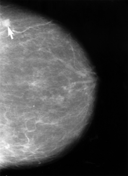
- Mammography is the most widely used test for screening and diagnosis. Abnormal findings: a mass, calcifications or architectural distortion. It is less useful in dense breasts and younger women.
- Ultrasound has similar sensitivity, suits younger or dense breasts and guides biopsy. MRI is the most sensitive study but is costly and less available.
- Findings are reported with BI-RADS categories 0 to 6, linking the image to a probability of cancer and a next step.
- Core needle biopsy (image-guided) is superior to fine needle aspiration and should be done whenever possible; a marker clip is placed at the site. Clinically positive nodes get an ultrasound-guided core biopsy.
- The biopsy must include ER, PR and HER2 testing. Routine staging scans are not needed for operable, asymptomatic early cancer; advanced disease gets CT chest–abdomen–pelvis with bone scan, or PET.
How is breast cancer staged under TNM and AJCC 8?
| Category | Definition |
|---|---|
| Tis | Carcinoma in situ; Paget disease without an underlying tumour |
| T1 | 2 cm or less (T1a 0.1–0.5 cm, T1b 0.5–1 cm, T1c 1–2 cm) |
| T2 | More than 2 cm up to 5 cm |
| T3 | More than 5 cm |
| T4a / T4b / T4c | Chest wall involvement / skin involvement / both |
| T4d | Inflammatory carcinoma |
| N1 | Mobile ipsilateral axillary nodes |
| N2 | Fixed or matted ipsilateral axillary nodes |
| N3a / N3b / N3c | Ipsilateral infraclavicular / internal mammary / supraclavicular nodes |
| M1 | Distant metastasis |

Stage groups follow: stage 0 is DCIS (non-invasive); stages I, IIA and IIB are early invasive cancer; IIIA, IIIB and IIIC are mostly locally advanced disease; stage IV is any metastatic cancer. Clinical staging is done before treatment; pathological staging after surgery.
When is breast-conserving surgery chosen over mastectomy?
Breast-conserving surgery (BCS) — lumpectomy or partial mastectomy with a margin of healthy tissue — can be offered to most patients with tumours under 5 cm if the breast is large enough for a good cosmetic result. BCS must be followed by whole-breast radiotherapy with a boost to the tumour bed to reduce local recurrence. Non-palpable lesions are localised before surgery with a wire or radioactive seed.
| Operation | What is removed | Main indications |
|---|---|---|
| Breast-conserving surgery | Tumour with a margin of normal tissue | Tumour under 5 cm with an adequate breast-to-tumour ratio; patient able to have radiotherapy |
| Simple (total) mastectomy | Whole breast with nipple–areola complex and pectoralis major fascia | Contraindication to BCS or patient preference |
| Modified radical mastectomy (MRM) | Simple mastectomy + axillary lymph node dissection | Mastectomy candidate with axillary disease |
| Radical (Halsted) mastectomy | Breast, axillary nodes and pectoral muscles | Seldom performed now |
- Contraindications to BCS / indications for mastectomy: large tumour, large tumour-to-breast ratio, chest wall or skin involvement, multifocal cancer, inflammatory cancer and inability to receive radiotherapy.
- Post-mastectomy radiotherapy is added in selected cases: tumour over 5 cm, chest wall or skin invasion, multifocal tumour, or 4 or more positive nodes.
- Axillary dissection preserves the long thoracic nerve and the thoracodorsal nerve.
- Complications of axillary dissection include lymphoedema, nerve injury, chronic pain and, rarely, lymphangiosarcoma.
What is sentinel lymph node biopsy and when is axillary dissection needed?
The sentinel node is the first node to receive lymph from the tumour. A radiotracer and/or blue dye is injected near the tumour or under the areola, and the 1–3 nodes that are hot or blue — usually in level I or II of the axilla — are removed. SLNB is the staging procedure for a clinically node-negative axilla in early invasive cancer, and it spares most women the morbidity of a full dissection.
| Situation | Axillary procedure |
|---|---|
| Clinically negative axilla, early cancer | Sentinel node biopsy |
| 1–3 positive sentinel nodes, no extranodal extension | No further axillary surgery needed |
| More than 3 positive nodes, or extranodal extension | Completion axillary dissection or axillary radiotherapy |
| Clinically positive axilla at diagnosis (after neoadjuvant therapy) | Axillary dissection |
| Bulky axillary disease or a large tumour with expected extensive nodal spread | SLNB not indicated |
How do tamoxifen, aromatase inhibitors and trastuzumab fit into treatment?
Endocrine therapy is indicated in all hormone receptor-positive patients and is given for 5 to 10 years; it reduces both recurrence and death. The choice depends mainly on menopausal status.
| Drug | Mechanism | Main use | Key adverse effects |
|---|---|---|---|
| Tamoxifen | SERM: oestrogen antagonist in breast, agonist in bone and endometrium; activated by CYP2D6 to endoxifen | HR-positive cancer, especially premenopausal women | Endometrial cancer, thromboembolism, hot flushes |
| Aromatase inhibitors (anastrozole, letrozole, exemestane) | Block conversion of androgens to oestrogen in peripheral tissues | Postmenopausal HR-positive cancer; in premenopausal women only with ovarian suppression | Arthralgia, bone loss and osteoporosis, hot flushes |
| Trastuzumab | Monoclonal antibody against HER2 | HER2-positive cancer (all HER2-positive tumours over 1 cm); often with pertuzumab | Cardiotoxicity (fall in LVEF), worse with anthracyclines |
About 17% of breast cancers overexpress HER2. Added to chemotherapy in early HER2-positive disease, trastuzumab cut the risk of recurrence by 52% and of death by 33% compared with chemotherapy alone. Other targeted options: CDK4/6 inhibitors (palbociclib) for HR-positive, HER2-negative tumours; pembrolizumab for triple-negative disease; PARP inhibitors for BRCA carriers. All triple-negative tumours over 1 cm receive chemotherapy, and neoadjuvant chemotherapy is increasingly used in triple-negative and HER2-positive early cancer and in locally advanced cancer (over 5 cm or clinically node-positive).