What are the dural venous sinuses?
The dural venous sinuses are blood-filled channels inside the cranial cavity that collect venous blood from the brain, meninges and skull and return it to the heart through the internal jugular veins. They lie between the outer periosteal and inner meningeal layers of the dura mater, mostly along the attached margins of the falx cerebri and tentorium cerebelli.
- They have no valves (the superior sagittal sinus has no muscular coat either), so blood — and infection or tumour cells — can flow in either direction.
- Their tributaries are the cerebral and cerebellar veins (including bridging veins crossing the subdural space), diploic veins of the skull bones, meningeal veins and emissary veins from the scalp.
- The superior sagittal sinus also receives cerebrospinal fluid through the arachnoid granulations.
- StatPearls counts seven major sinuses — superior sagittal, inferior sagittal, straight, transverse, sigmoid, cavernous and superior petrosal — and calls the cavernous sinus the most important clinically.
Which dural venous sinuses are paired and which are unpaired?
| Unpaired (midline) | Paired (one on each side) |
|---|---|
| Superior sagittal sinus | Transverse sinus |
| Inferior sagittal sinus | Sigmoid sinus |
| Straight sinus | Cavernous sinus |
| Occipital sinus | Superior petrosal sinus |
| Intercavernous sinus | Inferior petrosal sinus |
| — | Sphenoparietal sinus |
| — | Basilar venous plexus |
| Sinus | Location | Drains into |
|---|---|---|
| Superior sagittal | Attached (upper) border of the falx cerebri, from near the crista galli and foramen caecum backwards | Confluence of sinuses |
| Inferior sagittal | Lower free border of the falx cerebri | Joins the great cerebral vein to form the straight sinus |
| Straight | Formed where the inferior sagittal sinus meets the great cerebral vein | Confluence of sinuses (can drain into a transverse sinus) |
| Occipital | Small channel in the posterior part of the tentorium cerebelli, draining the occiput | Confluence of sinuses |
| Transverse | Attached margin of the tentorium cerebelli on the occipital bone | Sigmoid sinus |
| Sigmoid | S-shaped groove in the posterior cranial fossa | Internal jugular vein at the jugular foramen |
| Cavernous | Each side of the sella turcica, above and lateral to the sphenoid sinus | Superior and inferior petrosal sinuses |
| Superior petrosal | Runs back from the cavernous sinus | Sigmoid sinus |
| Inferior petrosal | Runs back and down from the cavernous sinus | Internal jugular vein |
How does venous blood drain from the brain to the internal jugular vein?
- Superior cerebral veins cross the subdural space as bridging veins into the superior sagittal sinus.
- The inferior sagittal sinus joins the great cerebral vein (of Galen) to form the straight sinus.
- The superior sagittal, straight and occipital sinuses meet at the confluence of sinuses (torcular Herophili) against the occipital bone.
- From the confluence, blood passes laterally into the right and left transverse sinuses.
- Each transverse sinus becomes the S-shaped sigmoid sinus.
- The sigmoid sinus leaves the skull through the jugular foramen as the internal jugular vein, which drains to the superior vena cava.
Superior sagittal sinus → confluence → transverse sinus → sigmoid sinus → internal jugular vein
The cavernous sinus reaches the same endpoint through the superior petrosal sinus (to the sigmoid sinus) and the inferior petrosal sinus (directly to the internal jugular vein).

What are the relations and contents of the cavernous sinus?
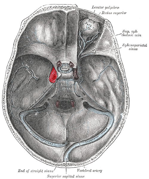
The cavernous sinus lies on each side of the sella turcica (and the pituitary), above and lateral to the sphenoid sinus. It extends from the superior orbital fissure in front to the apex of the petrous temporal bone behind. Inside, it is a network of trabeculated venous spaces — hence 'cavernous'. The two sinuses are joined by the intercavernous sinuses, which is how infection spreads from one eye to the other.
| Position | Structures | Clinical note |
|---|---|---|
| Within the sinus (lumen) | Internal carotid artery (with its sympathetic plexus) and the abducens nerve (VI) | CN VI runs free in the sinus, close to the inferolateral side of the ICA — it is the first nerve affected |
| Lateral wall (between the dural leaves) | Oculomotor (III), trochlear (IV), ophthalmic (V1) and maxillary (V2) nerves | Protected by dura, so they are involved later |
| Direction | Vessels |
|---|---|
| Receives (tributaries) | Superior and inferior ophthalmic veins, superficial middle cerebral vein, sphenoparietal sinus, middle meningeal vein |
| Communicates with | Pterygoid venous plexus (through emissary veins) and the facial vein (through the ophthalmic veins); the opposite cavernous sinus (intercavernous sinuses) |
| Drains into | Superior petrosal sinus (to the sigmoid sinus) and inferior petrosal sinus (to the internal jugular vein) |
What causes cavernous sinus thrombosis and how does it present?
Cavernous sinus thrombosis is rare but life-threatening. Most cases are septic: infection spreads through the valveless facial and ophthalmic veins or from neighbouring sinuses. The classic source is the 'danger triangle' of the face — the area from the corners of the mouth to the bridge of the nose (the nasolabial triangle). StatPearls lists sphenoid and ethmoid sinusitis as possibly the most common cause, along with orbital cellulitis, dental infection, otitis media and mastoiditis. Aseptic causes include trauma, surgery and pregnancy.
- Organism — Staphylococcus aureus in about two-thirds of cases (some methicillin-resistant), streptococci in about 20%; fungi (mucormycosis, aspergillosis) in immunocompromised patients.
- Symptoms — fever, headache (50–90%), periorbital swelling, eye pain, diplopia and visual loss; usually starts in one eye and spreads to the other.
- Eye signs in about 90%: chemosis, ptosis, proptosis, painful or restricted eye movements, papilloedema or retinal haemorrhages.
- Nerve palsies — CN VI palsy (failure of abduction) is the commonest and usually earliest; most cases progress to complete external ophthalmoplegia (III, IV, VI). Loss of V1/V2 sensation and a reduced corneal reflex; Horner syndrome may occur.
- Fever pattern — a 'picket fence' fever, with sharp spikes returning to normal.
- Blindness occurs in 8–15%.
Diagnosis is by contrast-enhanced CT or MRI with venography (CT venogram or contrast MR venogram); these show a widened cavernous sinus with a convex lateral wall, filling defects and thrombosed superior ophthalmic veins. Treatment is prolonged IV antibiotics — an anti-staphylococcal agent (vancomycin if MRSA is likely), a third-generation cephalosporin and metronidazole — usually for 3–4 weeks, with anticoagulation (unfractionated or low-molecular-weight heparin) recommended by most experts. Retrospective data suggest heparin lowers mortality from about 40% to 14%.
What are emissary veins and arachnoid granulations?
Emissary veins are valveless veins that pass through foramina in the skull and connect scalp (extracranial) veins with the dural venous sinuses and diploic veins. Blood normally flows inwards, but the direction can reverse to relieve raised intracranial pressure, and they help cool the brain. Because they are valveless, they are also a route for scalp and facial infection to reach the sinuses.
| Emissary vein | Connects |
|---|---|
| Parietal | Superficial temporal vein branches ↔ superior sagittal sinus |
| Mastoid | Posterior auricular or occipital vein ↔ transverse or sigmoid sinus |
| Occipital | Occipital vein ↔ transverse sinus |
| Posterior condylar | Internal vertebral plexus ↔ sigmoid, marginal or occipital sinus |
| Ophthalmic veins (through the superior orbital fissure) | Orbital veins ↔ cavernous sinus |
Arachnoid villi and granulations are outpouchings of the arachnoid into the venous sinuses that act as one-way valves, returning CSF from the subarachnoid space to the venous blood. The largest number lie in the walls of the superior sagittal sinus. When a sinus clots, CSF absorption fails and intracranial pressure rises — the reason papilloedema and headache are prominent in sinus thrombosis.

What are the risk factors and features of cerebral venous sinus thrombosis?
Cerebral venous sinus thrombosis (CVST) has an estimated incidence of 3–4 per million per year and is about three times commoner in women, mostly of childbearing age. At least one risk factor is found in over 85% of patients.
| Group | Examples |
|---|---|
| Oestrogen-related | Oral contraceptive pill, pregnancy and the puerperium, oestrogen-containing HRT, obesity |
| Inherited thrombophilia | Factor V Leiden, prothrombin G20210A, protein C, protein S or antithrombin deficiency, hyperhomocysteinaemia |
| Acquired prothrombotic states | Antiphospholipid syndrome, nephrotic syndrome, malignancy, myeloproliferative disorders (JAK2), SLE, inflammatory bowel disease |
| Local causes | Otitis media and mastoiditis (adjacent sigmoid and transverse sinuses), head injury, neurosurgery, jugular catheterisation, lumbar puncture |
| Other triggers | Dehydration, COVID-19, vaccine-induced thrombotic thrombocytopenia |
Two mechanisms explain the clinical picture: blocked cerebral veins raise venous and capillary pressure, causing venous infarction, oedema and haemorrhage; a blocked sinus stops CSF absorption, raising intracranial pressure.
- Headache in 80–90% — usually builds over days, worse with coughing or Valsalva, not relieved by sleep; sometimes thunderclap, mimicking subarachnoid haemorrhage.
- Raised ICP signs — papilloedema, diplopia from a CN VI palsy; isolated headache without other signs in up to 25%.
- Seizures in about 40% (often focal) — far commoner than in arterial stroke.
- Focal deficits in up to 44%, most often hemiparesis, which do not follow an arterial territory and may be bilateral.
- Straight-sinus thrombosis or a large haemorrhagic venous infarct can compress the diencephalon and brainstem, leading to coma and herniation.
- Onset is subacute (48 hours to 4 weeks) in nearly 60%.
How is venous sinus thrombosis diagnosed and treated, and what is the empty delta sign?
| Sign | Modality | What it shows |
|---|---|---|
| Dense triangle sign | Non-contrast CT | Triangular hyperdensity (fresh clot) in the superior sagittal sinus |
| Cord sign | Non-contrast CT | Curvilinear hyperdensity in a thrombosed cortical vein (visible for up to 2 weeks) |
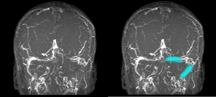
| Empty delta sign | Contrast-enhanced CT (also contrast MRI) | Triangular non-enhancing clot in the superior sagittal sinus outlined by enhancement from collateral venous flow |
| Filling defect / absent flow | CT venography, MRI with MR venography | Confirms the diagnosis; MRI + MRV is the gold standard |
- Non-contrast CT is the first scan, but sinus hyperdensity is seen in only about one-third of cases — so a normal CT does not exclude CVST.
- CT venography can be done immediately after the plain CT and performs comparably to MR venography.
- MRI with MRV is the most sensitive approach and shows parenchymal oedema well.
- A negative D-dimer does not exclude CVST (false negatives up to about 26%).
Treatment starts with control of raised intracranial pressure (head elevation, osmotherapy) and seizures, then anticoagulation as soon as the diagnosis is confirmed — even if there is intracerebral haemorrhage. Intravenous unfractionated heparin or subcutaneous LMWH is used first, then a vitamin K antagonist (target INR 2–3) or a direct oral anticoagulant such as dabigatran or rivaroxaban; LMWH is preferred in pregnancy. Duration is 3–6 months for provoked and 6–12 months for unprovoked thrombosis, and indefinite after recurrence or with severe thrombophilia. Catheter-directed therapy or decompression is reserved for selected patients who deteriorate.
How are the dural venous sinuses asked in NEET PG and INI-CET?
- Contents of the cavernous sinus — which nerve lies inside (VI) and which in the lateral wall (III, IV, V1, V2); 'not a content' answers: V3, optic nerve.
- First nerve affected in cavernous sinus thrombosis — abducens.
- Danger area of the face — spread to the cavernous sinus via valveless facial and ophthalmic veins.
- Drainage sequence — superior sagittal → confluence → transverse → sigmoid → internal jugular vein; inferior sagittal + great cerebral vein → straight sinus.
- Arachnoid granulations — mostly into the superior sagittal sinus.
- Empty delta sign — contrast CT sign of superior sagittal sinus thrombosis; dense triangle and cord signs on plain CT.
- CVST management — anticoagulate even when there is haemorrhage.
Practise with the NEET PG anatomy PYQs and INI-CET PYQs. Related pages: Horner syndrome, Virchow's triad and the Glasgow Coma Scale.