What is the inguinal canal and where does it lie?
The inguinal canal is a passage through the lower anterior abdominal wall, lying just above the inguinal ligament. It begins at the deep (internal) inguinal ring, runs medially and downwards obliquely through the layers of the abdominal wall, and ends at the superficial (external) inguinal ring. Radiopaedia gives its length as about 4 cm; StatPearls gives 4–6 cm in the adult, shorter in children.
It exists because the testis descends from the posterior abdominal wall to the scrotum, guided by the gubernaculum and preceded by a peritoneal pouch, the processus vaginalis. The canal is the path the testis and its cord take. Normally the processus closes after descent; if it stays open, peritoneal fluid can track down it (hydrocele) or bowel can follow it (indirect inguinal hernia). The same canal in the female carries the round ligament of the uterus.
Because it is a natural canal with two openings, it can widen and let abdominal contents through. A chronic rise in abdominal pressure is the main driver of these groin hernias, and repairing them well depends on knowing the canal's walls exactly.
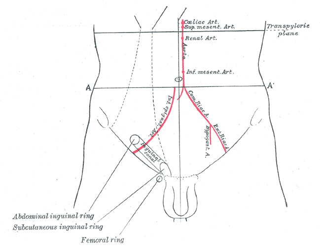
What forms the walls, roof and floor of the inguinal canal?
| Boundary | Formed by | Exam note |
|---|---|---|
| Roof (superior wall) | Arching fibres of internal oblique and transversus abdominis | The two Muscles |
| Anterior wall | External oblique aponeurosis, with internal oblique contribution | Two Aponeuroses; opened first in open repair |
| Floor (inferior wall) | Inguinal ligament, with the lacunar ligament medially | Two Ligaments; inguinal ligament runs ASIS → pubic tubercle |
| Posterior wall | Transversalis fascia, with the conjoint tendon medially | Tendon + fascia; the wall that fails in a direct hernia |
Conjoint tendon (inguinal falx, Henle's ligament): the common aponeurosis of the internal oblique and transversus abdominis, inserting into the pubic crest and pectineal line just deep to the superficial ring. It forms the main part of the medial posterior wall, directly behind the superficial ring — so it buttresses the ring against a direct push. Classic tissue repairs (Bassini, Shouldice, McVay) suture the conjoint tendon to strengthen this wall.
Where are the deep and superficial inguinal rings?
| Feature | Deep (internal) ring | Superficial (external) ring |
|---|---|---|
| Opening in | Transversalis fascia | External oblique aponeurosis |
| Shape | Round / oval | Triangular (V-shaped), edges held by intercrural fibres |
| Surface position | About 1 cm above the midpoint of the inguinal ligament | Just above and lateral to the pubic tubercle |
| Relation to inferior epigastric vessels | Lateral to them | Medial — sits in front of the conjoint tendon |
| Wall layer → cord covering | Transversalis fascia → internal spermatic fascia | External oblique → external spermatic fascia |
| Hernia that enters here | Indirect inguinal hernia | Both indirect and (sometimes) direct hernias exit here |
What passes through the inguinal canal in males and females?
| Male | Female | Both sexes |
|---|---|---|
| Spermatic cord and its contents | Round ligament of the uterus | Ilioinguinal nerve (usually outside the cord, on its front) |
| — | — | Genital branch of the genitofemoral nerve (enters through the deep ring) |
- Spermatic cord contents: vas deferens, testicular artery, artery to the vas, cremasteric artery, the pampiniform plexus of veins, lymphatics and the genital branch of the genitofemoral nerve.
- Coverings of the cord pick up one layer from each wall it passes: external spermatic fascia (from external oblique), cremasteric muscle and fascia (from internal oblique and transversus abdominis), internal spermatic fascia (from transversalis fascia).
- Deep to superficial around the testis: tunica vaginalis → internal spermatic fascia → cremasteric fascia → external spermatic fascia → dartos.
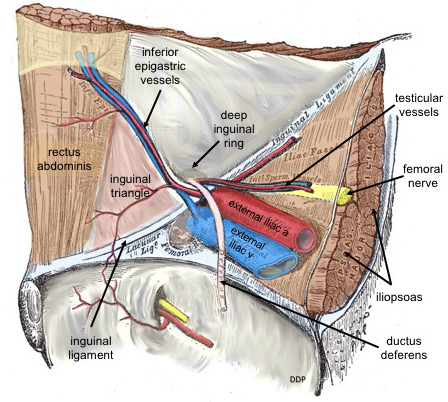
What are the boundaries of Hesselbach's triangle?
Hesselbach's (inguinal) triangle is the weak area of the posterior wall of the canal, seen from inside the abdomen. Direct inguinal hernias come through it.
| Border | Structure |
|---|---|
| Lateral (superolateral) | Inferior epigastric vessels |
| Medial | Lateral border of rectus abdominis (rectus sheath) |
| Inferior (base) | Inguinal ligament |
The conjoint tendon reinforces the medial part of the triangle. A weakness here in young athletes is described as a Bugosa or Gill-Ogilvie hernia (a type of 'sports hernia'); in older adults the posterior wall simply weakens with age and strain.
How does a direct inguinal hernia differ from an indirect one?
| Feature | Indirect | Direct |
|---|---|---|
| Route | Through the deep ring along the canal | Straight through the posterior wall (Hesselbach's triangle) |
| Relation to inferior epigastric vessels | Lateral | Medial |
| Cause | Patent processus vaginalis (congenital) | Weak abdominal wall — age, strain, previous surgery |
| Typical patient | Children and young adults; any age | Older adults |
| Frequency | Commonest — about twice as common as direct; commonest groin hernia in both sexes | Less common |
| Side | More often right (later closure of the right processus vaginalis) | — |
| Reaches the scrotum | Yes, can descend into the scrotum (lies anterior to the cord) | Usually stays in the groin |
Epidemiology: the lifetime risk of an inguinal hernia is about 27% in men and 3% in women, and inguinal hernias are 9 to 12 times commoner in males. Risk rises with age and with a positive family history.

How is a femoral hernia distinguished from an inguinal hernia?
| Feature | Inguinal hernia | Femoral hernia |
|---|---|---|
| Neck of sac | Above the inguinal ligament | Below the inguinal ligament, through the femoral ring |
| Relation to pubic tubercle | Emerges at the superficial ring, above the ligament | Below and lateral to the tubercle |
| Sex | Much commoner in men | About 4 times commoner in women |
| Commonest groin hernia in women? | Yes — still the commonest in women | No |
| Strangulation | Lower risk | Highest of all hernias — about 15–20% |
| Management | Elective repair (mesh) | Repair at diagnosis; emergency if obstructed |
Femoral ring boundaries: inguinal ligament in front (anterosuperiorly), Cooper's (pectineal) ligament behind, femoral vein laterally and the lacunar ligament medially. These unyielding borders around a small ring help explain why femoral hernias carry the highest strangulation rate and are repaired as soon as they are found.

Which nerves are at risk during inguinal hernia surgery?
| Nerve | Root | Course | Supplies |
|---|---|---|---|
| Ilioinguinal | L1 | In the canal on the front of the cord; exits the superficial ring | Skin of upper medial thigh, root of penis and anterior scrotum (mons and labia majora in females) |
| Genital branch of genitofemoral | L1–L2 | Enters through the deep ring with the cord | Cremaster (motor) and scrotal/labial skin |
| Iliohypogastric | L1 (often with T12 branches) | Pierces the muscles above the canal — not in it | Suprapubic skin |
In open repair, StatPearls stresses identifying and protecting the ilioinguinal, genitofemoral and iliohypogastric nerves; the ilioinguinal nerve and the genital branch can be compressed by sutures or mesh near the internal oblique, giving chronic groin pain or tingling over their territory. In laparoscopic repair (TEP/TAPP), tacks placed below the iliopubic tract can injure the genitofemoral or lateral femoral cutaneous nerve. The genital branch of the genitofemoral nerve supplies the cremaster, and both it and the ilioinguinal nerve feed sensation into the cremasteric reflex.
Repairs in one line: the accepted standard open repair is the Lichtenstein tension-free flat mesh, sutured to the inguinal ligament and the conjoint tendon area, with a keyhole for the cord at a new internal ring; tissue repairs (Bassini, Shouldice, McVay) failed more often because of tension. McVay (Cooper's ligament) repair is the tissue repair that also closes the femoral space.
What are the high-yield exam traps on the inguinal canal?
- Deep ring = gap in transversalis fascia; superficial ring = gap in external oblique aponeurosis.
- Floor = inguinal + lacunar ligaments; posterior wall = transversalis fascia + conjoint tendon (medially).
- Inferior epigastric vessels: indirect lateral, direct medial.
- Hesselbach: rectus (medial), inferior epigastrics (lateral), inguinal ligament (base).
- Femoral hernia: below and lateral to the pubic tubercle; highest strangulation risk.
- Ilioinguinal nerve runs in the canal but does not pass through the deep ring and is not inside the cord.
- Internal spermatic fascia ← transversalis fascia; cremaster ← internal oblique and transversus; external spermatic fascia ← external oblique.
Related reading: the pelvic peritoneal recess in pouch of Douglas, and nerve-supply tables in hand muscles and nerve supply. For past papers, see NEET PG anatomy PYQs and NEET PG surgery PYQs.