Which nerves are injured in the lower limb and what do they cause?
The lower limb is supplied by the lumbar plexus (anterior rami of L1 to L4: obturator, femoral, lateral femoral cutaneous) and the sacral plexus (sciatic nerve and its two divisions, the gluteal nerves). Most exam questions ask you to match a lesion to its clinical deficit, so it helps to learn one pattern per nerve.
| Nerve (roots) | Typical site of injury | Main deficit |
|---|---|---|
| Common peroneal / fibular (L4–S2) | Fibular neck; knee dislocation; casts; leg crossing | Foot drop, weak eversion, numbness over dorsum of foot and lateral leg |
| Sciatic (L4–S3) | Hip dislocation or surgery; deep gluteal compression | Weak knee flexion and all ankle movements; sensory loss over most of the foot |
| Superior gluteal (L4, L5, S1) | Hip surgery or fracture; gluteal intramuscular injection | Trendelenburg gait (weak gluteus medius and minimus) |
| Femoral (L2–L4) | Pelvic haematoma, lithotomy position, hip surgery | Quadriceps wasting, loss of knee extension; numb anterior thigh and medial leg |
| Obturator (L2–L4) | Pelvic surgery; obturator hernia | Weak thigh adduction; small patch of medial thigh numbness |
| Lateral femoral cutaneous (L2, L3) | Under the inguinal ligament | Painful, purely sensory numbness of the anterolateral thigh |
| Tibial / posterior tibial (L4–S3) | Tarsal tunnel behind the medial malleolus | Burning pain and numbness of the sole; worse on walking |

What happens when the common peroneal nerve is injured?
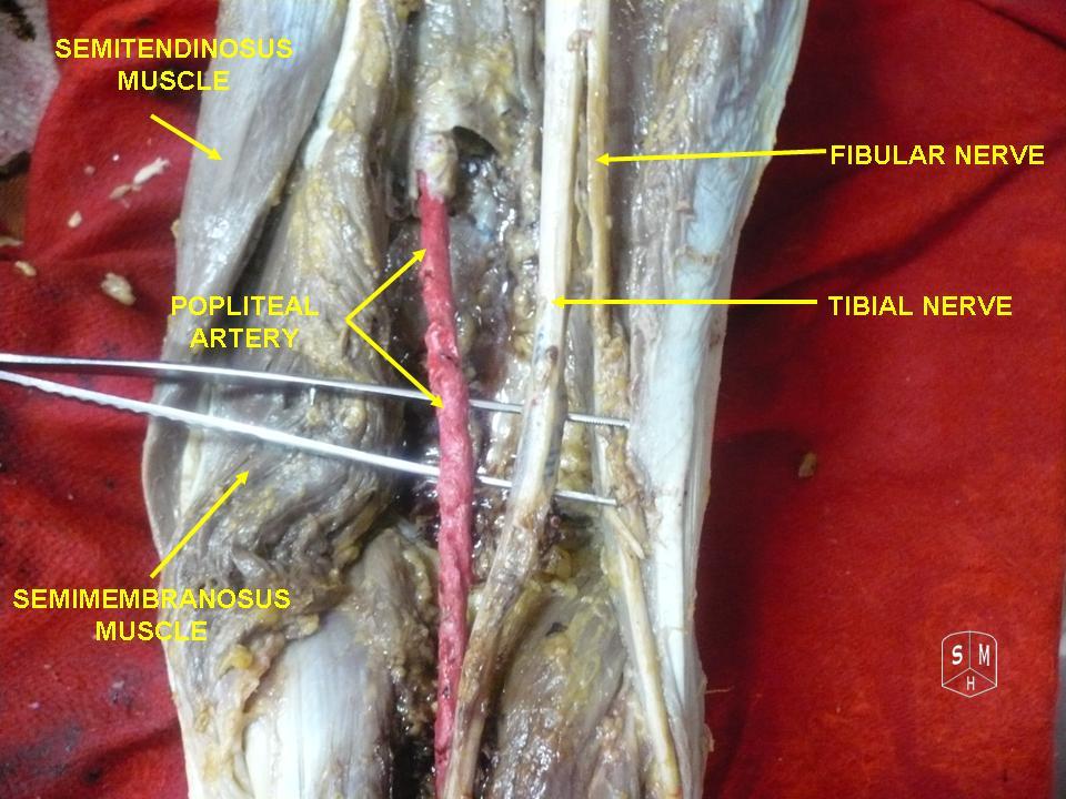
The common peroneal (fibular) nerve is one of the two terminal branches of the sciatic nerve, which divides into the tibial and common peroneal nerves just above the popliteal fossa. Its fibres come from the posterior divisions of L4–S2. It runs behind the biceps femoris, winds around the neck of the fibula where it lies subcutaneously, and then divides into the deep and superficial peroneal nerves.
| Branch | Motor | Sensory |
|---|---|---|
| Deep peroneal | Dorsiflexion of the ankle; extension of the great toe | Skin of the first web space between the first and second toes |
| Superficial peroneal | Eversion of the foot | Dorsum of the foot, except the first web space |
Peroneal neuropathy is the most common compressive neuropathy of the lower extremity and, after median and ulnar neuropathies, the most common focal neuropathy. A classical presentation is foot drop: weakness of ankle and toe dorsiflexion plus weak eversion, so the patient catches the toes while walking and lifts the knee high — the high-steppage gait — with numbness over the lateral leg and dorsum of the foot.
| Category | Examples |
|---|---|
| Trauma at the knee | Knee dislocation (up to 40% of acute dislocations injure the nerve), direct blow or laceration at the fibular head, proximal fibula or tibial plateau fracture |
| External compression | Tight plaster or splint, compression bandage, habitual leg crossing, prolonged bed rest, poorly padded fibular head during anaesthesia or surgery |
| Anatomical | Fibrous band at the origin of peroneus longus; fabella (a sesamoid in gastrocnemius) pressing on the nerve |
| Mass lesions | Intraneural ganglion cyst, peripheral nerve tumours |
| Behavioural | Athletes and workers who kneel or squat for long periods |
What causes foot drop and how do you tell the causes apart?
Foot drop is weakness of the dorsiflexors, producing a high-steppage walking pattern. It is a sign, not a diagnosis. A lesion of the L5 root, lumbar plexus, sciatic nerve, common peroneal nerve or deep peroneal nerve can cause it, plus muscle disease and central causes.
| Level | What else is involved | Clues |
|---|---|---|
| L5 radiculopathy (the commonest radicular cause) | Dorsiflexors and evertors; pain radiating from the lumbar region down the posterior thigh, anterolateral leg and big toe | Back pain, disc herniation; sensory symptoms on the medial foot and first web space |
| Lumbosacral plexopathy | Hip girdle weakness — gluteus medius (abduction) and gluteus maximus (extension) | Resembles sciatic neuropathy plus proximal weakness |
| Sciatic neuropathy | Plantar flexors (gastrocnemius, soleus), inversion, hamstrings; whole-foot sensory loss ('flail foot') | Hip injury or surgery. Incomplete lesions often mimic a peroneal palsy because the peroneal fascicles are more vulnerable |
| Common peroneal neuropathy | Dorsiflexion and eversion only; inversion and plantar flexion preserved | Fibular-neck compression, Tinel sign at the neck |
| Mononeuritis multiplex | Asymmetric painful multifocal nerve loss; the sciatic nerve is often involved | Leprosy, vasculitis, HIV, hepatitis, rheumatoid arthritis |
| Charcot-Marie-Tooth disease | Bilateral foot drop with wasting of lower leg muscles ('stork leg' appearance) | Inherited demyelinating neuropathy; family history |
Management of peroneal foot drop is usually conservative: an ankle-foot orthosis (AFO), physiotherapy and removal of the cause (loosening a cast, stopping leg crossing). Surgery is considered for a rapidly deteriorating lesion, no improvement after about 3 months, or an open injury with suspected laceration, which should be explored and repaired within 72 hours. Foot drop after knee dislocation has a poorer prognosis, and tendon transfers are often needed for refractory palsy.
What are the effects of sciatic and gluteal nerve injuries?
The sciatic nerve (L4–S3) is the largest nerve of the body. It leaves the pelvis through the greater sciatic foramen below the piriformis and splits in the popliteal fossa into the tibial (medial) and common fibular (lateral) divisions; the tibial division also supplies the hamstring part of adductor magnus, semitendinosus, semimembranosus and the long head of biceps. In the commonest anatomical variant the nerve leaves undivided below the piriformis, but in some people the fibular division passes through the muscle and can be compressed there.
- Sciatic neuropathy (most often from traumatic hip injury or surgery) is the second most common mononeuropathy of the lower limb and usually causes foot drop with loss of plantar flexion and inversion, sensory loss over the entire foot and weak knee flexion.
- Sciatica is a different entity — buttock-to-leg pain from nerve root irritation, most often a disc herniation at L5 or S1, not a peripheral nerve injury.
- The superior gluteal nerve (L4, L5, S1) leaves the pelvis above the piriformis — the only nerve to do so — and supplies gluteus medius, gluteus minimus and tensor fasciae latae.
- It can be injured by hip dislocation or fracture, hip arthroplasty and intramuscular injection into the buttock.
What do femoral and obturator nerve injuries cause?
| Feature | Femoral nerve | Obturator nerve |
|---|---|---|
| Roots | Dorsal divisions of L2–L4 | L2–L4 (lumbar plexus) |
| Motor | Iliacus, pectineus, quadriceps (hip flexion, knee extension) | Adductors of the thigh (adductor longus, brevis, magnus, gracilis, obturator externus) |
| Sensory | Anterior and medial thigh; saphenous branch to the medial leg and foot | Skin of the medial upper thigh; articular branches to hip and knee |
| Injury | Pelvic haematoma or abscess in psoas/iliacus; hip and abdominal surgery; lithotomy position compressing the nerve under the inguinal ligament; femoral IM nail screws | Pelvic surgery (prolapse repair, laparoscopic lymphadenectomy); obturator hernia; entrapment in athletes |
| Clinical picture | Quadriceps wasting, loss of knee extension, weaker hip flexion, loss of patellar reflex; the patient may fall on standing after delivery | Weak adduction; sudden adductor jerk during transurethral bladder-tumour resection |
For the femoral nerve, the nerve lies between the psoas major and iliacus, so a growing haematoma or abscess there compresses it. Most patients recover satisfactorily, though some need nerve repair or grafting. For the obturator nerve, an obturator hernia typically presents in elderly, thin women or patients with raised intra-abdominal pressure and causes small-bowel obstruction; entrapment in athletes causes exercise-induced medial thigh pain.
What is meralgia paraesthetica?
Meralgia paraesthetica (Bernhardt-Roth syndrome) is compression of the lateral femoral cutaneous nerve (LFCN) — a pure sensory nerve from the posterior divisions of L2 and L3. It crosses the iliacus toward the anterior superior iliac spine and passes under, through or over the inguinal ligament, where it is most vulnerable. The anterior branch supplies the anterior thigh to the knee and the posterior branch the lateral thigh to the greater trochanter.
| Aspect | Details |
|---|---|
| Symptoms | Burning pain, paraesthesia and reduced sensation over the upper lateral thigh; usually unilateral; no motor loss, reflexes and straight-leg raise normal |
| Triggers | Prolonged hip extension (walking, standing); relieved by sitting (hip flexion); patients rub the thigh and may lose hair there |
| Causes | Tight belts, seat belts or clothing; obesity, pregnancy, abdominal tumours; diabetes, alcohol, lead poisoning, hypothyroidism; hip, spine, iliac-crest graft and laparoscopic surgery |
| Test | Pelvic compression test (side-lying, downward pressure on the ilium for about 45 seconds) — positive if symptoms ease; reported sensitivity 95% and specificity 93% |
| Treatment | Reassurance, avoid tight garments, weight loss; NSAIDs, topical lidocaine or capsaicin; gabapentin or carbamazepine if persisting beyond 1–2 months; nerve block; surgery (decompression or transection) only for refractory cases |
| Prognosis | About 85% recover spontaneously with conservative care; pregnancy-related cases improve after delivery |
What is tarsal tunnel syndrome?
Posterior tarsal tunnel syndrome is an entrapment of the tibial nerve in the fibro-osseous tunnel behind and below the medial malleolus. The roof is the flexor retinaculum (laciniate ligament, running from the tip of the medial malleolus to the medial calcaneus); the floor is formed by the medial tibia, talus and calcaneus. It is often compared with carpal tunnel syndrome but is considerably less common.
| Order | Structure |
|---|---|
| 1 | Tibialis posterior tendon |
| 2 | Flexor digitorum longus tendon |
| 3 | Posterior tibial artery and vein |
| 4 | Posterior tibial nerve (L4–S3) |
| 5 | Flexor hallucis longus tendon |

Within the tunnel the posterior tibial nerve divides into the medial plantar nerve (sensation to the medial foot and plantar first three and a half toes; abductor hallucis, flexor digitorum brevis, flexor hallucis brevis and first lumbrical) and the lateral plantar nerve (plantar lateral one and a half toes; most intrinsic muscles). The medial calcaneal nerve to the heel usually arises before the tunnel.
- Symptoms: medial ankle pain, plantar dysaesthesia (burning, tingling), worse with walking; sensory symptoms outweigh weakness.
- Risk factors: a history of ankle fracture or sprain (17–43% of patients), runners with hyperpronation, diabetes, hypothyroidism, gout, mucopolysaccharidosis, hyperlipidaemia, and mass lesions such as tibial nerve schwannoma.
- Diagnosis: clinical, supported by electrodiagnostic studies and imaging for structural causes.
- Anterior tarsal tunnel syndrome is a separate, rare entity: the deep peroneal nerve is compressed under the extensor retinaculum against the navicular and talus.
How are lower limb nerve injuries tested in the exam?
- Foot drop with eversion weakness and dorsal foot numbness — common peroneal nerve at the fibular neck (the commonest answer).
- Foot drop after hip surgery with plantar flexion and inversion weakness — sciatic nerve; after a knee dislocation — common peroneal nerve.
- Hip sags on the opposite side during stance — superior gluteal nerve; check for gluteal injection or hip surgery.
- Quadriceps wasting, absent knee jerk — femoral nerve; remember the saphenous branch for the medial leg.
- Adductor weakness — obturator nerve; adductor jerk during TURBT is an anaesthetic complication.
- Numb outer thigh in an obese or pregnant patient — lateral femoral cutaneous nerve (meralgia paraesthetica).
- Burning sole, medial ankle — tarsal tunnel; know the order of structures under the flexor retinaculum.