How do you localise an upper limb nerve injury?
Every peripheral nerve gives off branches in a fixed order. A lesion knocks out every branch distal to it and spares everything above. So the examiner's question — where is the lesion? — is answered by finding the highest muscle that is weak and the highest sensory branch that is lost.
- Name the nerve from the pattern — wrist drop (radial), claw (ulnar), ape thumb or failed OK sign (median), flat shoulder with failed abduction (axillary).
- Check the 'marker' muscle supplied just above the suspected level — triceps for the radial nerve (axilla vs spiral groove), flexor carpi ulnaris and the ulnar half of FDP for the ulnar nerve (elbow vs wrist), FDS and pronators for the median nerve (elbow vs wrist).
- Check the 'marker' skin branch — the palmar cutaneous branch of the median nerve and the dorsal cutaneous branch of the ulnar nerve both leave above the wrist, so their skin is spared in wrist lesions.
- Look for root-level clues — Horner syndrome, scapular winging or weakness spanning several nerves point to the brachial plexus.
What do brachial plexus lesions look like?
The brachial plexus is formed by the ventral rami of C5 to T1 and ends in five terminal nerves — musculocutaneous, axillary, radial, median and ulnar. Injuries are usually traction injuries: the head and neck forced away from the shoulder damage the upper trunk, and hyperabduction of the arm damages the lower trunk.
| Lesion | Mechanism | Key deficit |
|---|---|---|
| Upper trunk (C5–C6) — Erb | Head and neck pulled away from the shoulder (birth injury; road accidents, mostly motorcycles) | Arm adducted, internally rotated, forearm pronated — 'waiter's tip'. Details on Erb's palsy |
| Lower trunk (C8–T1) — Klumpke | Hyperabduction traction of the arm, including during birth | Total claw hand (all intrinsic muscles), sensory loss on the medial forearm and hand; Horner syndrome if the T1 sympathetic outflow is hit |
| Long thoracic nerve (C5–C7) | Injury anywhere along its course | Paralysed serratus anterior → medial scapular winging |
What happens in axillary nerve injury?
The axillary nerve (posterior cord, C5–C6) leaves the axilla through the quadrangular space — bounded by teres minor above, teres major below, the long head of triceps medially and the surgical neck of the humerus laterally. It supplies deltoid and teres minor and, through the superior lateral cutaneous nerve of the arm, the skin over the lower deltoid.
| Cause | Motor loss | Sensory loss |
|---|---|---|
| Anterior shoulder dislocation (the commonest nerve injury with dislocation); fracture of the surgical neck; quadrangular space syndrome in overhead athletes; shoulder surgery | Deltoid → weak abduction and shoulder weakness; teres minor → weak external rotation | 'Regimental badge' area over the lateral shoulder |

How does the level of radial nerve injury change the deficit?
The radial nerve (posterior cord) supplies triceps in the arm, brachioradialis and the wrist and finger extensors. Near the lateral epicondyle it splits into the purely motor posterior interosseous nerve (PIN) and the purely sensory superficial radial nerve. Its classic sign is wrist drop — obvious when the forearm is pronated, easily missed when the hand is supinated and gravity helps extension.
| Level | Typical cause | Triceps | Wrist drop | Sensory loss |
|---|---|---|---|---|
| Axilla | Crutch palsy, shoulder dislocation | Weak | Yes | Posterior arm and forearm + dorsoradial hand |
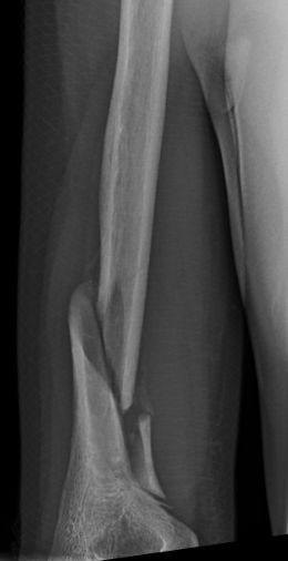
| Spiral groove (mid-humerus) — commonest site | Humeral shaft fracture; Saturday night palsy (arm compressed during deep sleep) | Spared | Yes; brachioradialis weak | Dorsum of hand, first web space side |
| Below the elbow — PIN | Entrapment at the arcade of Frohse in supinator; elbow fractures or dislocation | Spared | Finger and thumb drop with only partial wrist weakness | None — PIN has no skin branch |
| Superficial radial nerve at the wrist (Wartenberg syndrome) | Tight wristbands, casts, handcuffs; repetitive pronation–supination | Spared | No | Dorsoradial hand only |
How does a median nerve injury at the elbow differ from one at the wrist?
The median nerve supplies no muscle in the arm. In the forearm it supplies pronator teres, flexor carpi radialis, palmaris longus and FDS, and through the anterior interosseous nerve (AIN) the lateral half of FDP, flexor pollicis longus and pronator quadratus. In the hand it supplies the thenar muscles (abductor pollicis brevis, opponens pollicis, superficial head of flexor pollicis brevis) and gives skin to the palmar thumb, index, middle and radial half of the ring finger.
| Level | Typical cause | Motor deficit | Sensory deficit |
|---|---|---|---|
| At or above the elbow (high lesion) | Supracondylar humerus fracture; ligament of Struthers / supracondylar process; between the heads of pronator teres | Forearm pronation weak; no PIP flexion of index and middle (FDS); no FPL; thenar paralysis → ape hand | Lateral 3½ digits and the thenar eminence |
| Anterior interosseous nerve | Usually part of complex trauma | FPL + lateral FDP + pronator quadratus → cannot make an 'OK' sign (pinch) | None — AIN has no cutaneous branch |
| At the wrist (carpal tunnel) | Carpal tunnel syndrome, wrist lacerations | Thenar weakness and wasting, loss of opposition → ape thumb | Lateral 3½ digits; thenar skin spared (palmar cutaneous branch leaves above the tunnel) |
Carpal tunnel syndrome is the commonest entrapment neuropathy of the upper limb (about 3% of the population; peak age 45–54; associations include pregnancy, hypothyroidism, diabetes and rheumatoid arthritis). Phalen test — wrists held fully flexed back-to-back for 1 minute reproduces symptoms; Tinel test — tapping over the tunnel (sensitivity about 50%, specificity about 77%). The benediction sign is linked by different authors to median or ulnar lesions; see the companion hand page for how it is taught.
How does an ulnar nerve injury at the elbow differ from one at the wrist?
The ulnar nerve (medial cord, C8–T1) passes behind the medial epicondyle and through the cubital tunnel under Osborne's ligament, then supplies flexor carpi ulnaris and the ulnar half of FDP (DIP flexion of ring and little fingers). At the wrist it enters Guyon's canal and supplies the hypothenar muscles, interossei, 3rd and 4th lumbricals, adductor pollicis and the deep head of flexor pollicis brevis. Its dorsal cutaneous branch leaves in the forearm to supply the dorsal ulnar hand.
| Level | Typical cause | Motor deficit | Sensory deficit |
|---|---|---|---|
| Elbow (cubital tunnel) — commonest entrapment site | Cubital tunnel syndrome (compression under Osborne's ligament) | FCU (wrist deviates radially on flexion) + ulnar FDP + all ulnar intrinsics | Palmar and dorsal ulnar 1½ fingers and hypothenar area |
| Guyon's canal zone 1 (before the split) | Ganglion, hook of hamate fracture | Intrinsics (FCU and FDP spared) | Palmar little and ring fingers; dorsum spared |
| Guyon's canal zone 2 | Ganglion, hook of hamate fracture | Pure motor — deep branch only | None |
| Guyon's canal zone 3 | Ulnar artery thrombosis, compression | None | Pure sensory — superficial branch |
- Froment's sign — holding paper between thumb and index finger; the thumb IP joint flexes (FPL, median) to compensate for a weak adductor pollicis.
- Wartenberg's sign — the little finger drifts into abduction because the palmar interossei are weak.
- Claw hand — hyperextension at the 4th and 5th MCP joints with IP flexion. A wrist lesion claws more than an elbow lesion, because an elbow lesion also weakens the ulnar FDP that flexes the IP joints (the ulnar paradox — see the hand page).
- Tinel's sign at the cubital tunnel or Guyon's canal, and symptoms reproduced by elbow flexion, support a compressive lesion.
What is the one-table summary of upper limb nerve lesions?
| Nerve / level | Classic cause | Sign to remember |
|---|---|---|
| Upper trunk C5–C6 | Traction (birth, motorcycle accidents) | Waiter's tip |
| Lower trunk C8–T1 | Hyperabduction, Pancoast tumour | Total claw ± Horner |
| Long thoracic | Nerve injury (C5–C7) | Medial scapular winging |
| Axillary | Anterior dislocation, surgical neck fracture | Lost abduction, regimental badge anaesthesia |
| Radial — axilla | Crutch palsy | Wrist drop + weak triceps |
| Radial — spiral groove | Shaft fracture, Saturday night palsy | Wrist drop, triceps spared |
| PIN | Arcade of Frohse | Finger drop, no sensory loss |
| Median — elbow | Supracondylar fracture | Weak FDS/FPL, ape hand, numb thenar skin |
| AIN | Complex forearm trauma | Cannot make OK sign, no sensory loss |
| Median — wrist | Carpal tunnel syndrome | Ape thumb, thenar skin spared |
| Ulnar — elbow | Cubital tunnel | Claw + weak FCU/FDP, dorsal hand numb |
| Ulnar — wrist | Guyon's canal | Marked claw, dorsal hand spared |

How are upper limb nerve injuries asked in NEET PG and INI-CET?
- Injury → nerve pairs: surgical neck fracture or anterior dislocation → axillary; mid-shaft or distal-third spiral humerus fracture → radial; supracondylar fracture → median; cubital tunnel compression → ulnar.
- Triceps spared + wrist drop → spiral groove; triceps weak → axilla.
- Finger drop without sensory loss → posterior interosseous nerve.
- Cannot make an O with thumb and index, sensation normal → anterior interosseous nerve.
- Numb fingers, normal thenar skin → carpal tunnel; numb dorsal ulnar hand → lesion above the wrist.
- Pure motor ulnar deficit → Guyon's canal zone 2.
- Claw hand + Horner → C8–T1 root (Klumpke).