What are pyodermas and how are they classified?
Pyoderma simply means a pus-producing skin infection. In the NEET PG and INI-CET syllabus the term covers the common bacterial infections of the skin and its appendages, nearly all caused by *Staphylococcus aureus* and/or *Streptococcus pyogenes* (group A beta-haemolytic streptococcus, GABHS). The useful way to learn them is by depth in the skin: the more superficial the infection, the more benign it is.
| Level | Condition | Typical organism | Key feature |
|---|---|---|---|
| Epidermis (superficial) | Impetigo (non-bullous, bullous) | S. aureus ± GABHS | Honey-coloured crust or flaccid bullae |
| Epidermis, toxin at distance | Staphylococcal scalded skin syndrome (SSSS) | Exfoliative-toxin-producing S. aureus | Widespread superficial peeling, Nikolsky positive |
| Superficial hair follicle | Folliculitis | S. aureus (commonest) | Follicular pustule on a red base |
| Deep hair follicle | Furuncle (boil) | S. aureus | Tender, red, fluctuant nodule |
| Several contiguous follicles | Carbuncle | S. aureus, often MRSA | Nodule with multiple draining sinuses, systemic symptoms |
| Deep epidermis into dermis | Ecthyma | S. aureus / GABHS | Punched-out ulcer with crust |
What are the types of impetigo and who gets them?
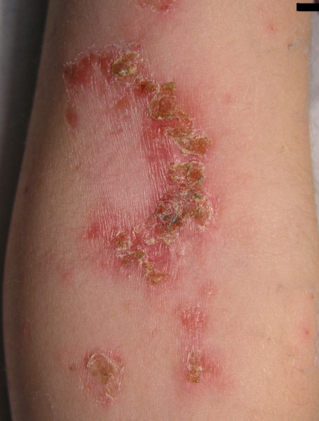
Impetigo is a common, highly contagious infection of the superficial layers of the epidermis. It is a disease of children, especially in hot, humid climates, and accounts for roughly 10% of skin complaints in the paediatric population. It is commonest at 2–5 years of age. It may be non-bullous or bullous, and either type can arise on previously normal skin (primary impetigo) or on skin already damaged by a wound, insect bite, burn, varicella, atopic dermatitis or scratching (secondary impetigo).
Bacteria need an entry point: once the skin barrier is breached, S. aureus and GABHS bind fibronectin receptors to colonise it. Overcrowding, daycare attendance, malnutrition, diabetes, immunosuppression and poor hygiene all raise the risk, and self-inoculation to new sites is very common.
| Feature | Non-bullous (impetigo contagiosa) | Bullous impetigo | Ecthyma |
|---|---|---|---|
| Share of cases | Commonest form | Much less common; >90% of cases in children under 2 years | Complication / deep form |
| Organism | S. aureus about 80%; GABHS about 10%; both about 10% | Almost exclusively S. aureus | S. aureus / GABHS |
| Lesion | Vesicle or pustule that ruptures → honey-coloured crust on a red base | Small vesicles → flaccid bullae with clear then cloudy fluid; no honey-coloured crust | Punched-out ulcer with violaceous margin, brown-black crust |
| Site | Face (perioral, nasal) and extremities | Intertriginous areas and trunk; may involve buccal mucosa | Deeper than ordinary impetigo; may follow bullous impetigo |
| Surrounding skin | Erythematous | Little erythema or oedema | Erythematous rim |
| Fever, nodes | Fever usually absent; mild regional lymphadenopathy | Fever more common; regional nodes usually absent | Variable |
Why does bullous impetigo form bullae?
In bullous impetigo the S. aureus at the lesion produces exfoliative toxin A, which causes loss of cell adhesion in the superficial epidermis. The result is a thin-walled, flaccid bulla that ruptures easily, leaving an erythematous base with a rim of scale. Importantly, in bullous impetigo the toxin acts locally, at the site of the infection. This is the contrast with SSSS, where the same type of toxin spreads through the blood.
How is impetigo diagnosed and treated?
Impetigo is a clinical diagnosis. Culture is reserved for suspected MRSA, outbreaks or lesions that fail to respond. The ASO response is weak after skin infection, so ASO titres are not used to diagnose impetigo, although streptococcal serology may help if post-streptococcal glomerulonephritis is suspected. Previously healthy adults with bullous impetigo should be considered for HIV testing.
- Localised, uncomplicated non-bullous impetigo: remove crusts with soap and water, then topical antibiotic alone — mupirocin, retapamulin or fusidic acid are the agents of choice.
- Systemic antibiotics for all bullous impetigo, and for non-bullous impetigo with more than five lesions, deep-tissue involvement, systemic signs, lymphadenopathy or oral-cavity lesions.
- Preferred oral agents: beta-lactamase-resistant drugs such as cephalexin (cephalosporins), dicloxacillin or amoxicillin-clavulanate. If cultures show streptococci only, oral penicillin is preferred.
- MRSA or high local MRSA prevalence: clindamycin or doxycycline. Trimethoprim-sulfamethoxazole acts on MRSA but does not reliably cover streptococci, so use it only when GABHS is not the cause or with an anti-streptococcal drug.
- Recurrent impetigo: look for carriage; the nose is the common reservoir and nasal mupirocin can be used for carriers.
Untreated impetigo usually heals in 14–21 days (about 20% resolve spontaneously); with treatment, cure is expected within about 10 days. Scarring is rare, though pigment change may occur. Children should avoid daycare or school for at least 24–48 hours after starting antibiotics.
What is staphylococcal scalded skin syndrome (SSSS)?
SSSS (also called Ritter disease or pemphigus neonatorum) is a dangerous toxin-mediated skin infection caused by certain S. aureus strains. The organism sits at a distant focus — nasopharynx, conjunctiva, umbilicus, ear or an abscess — and releases exfoliative (epidermolytic) toxins A and B. These are serine proteases that travel in the blood and cleave desmoglein-1 (Dsg1), the desmosomal cadherin that holds keratinocytes together in the stratum granulosum. The split therefore lies just beneath the stratum corneum.
Because the toxin, not the bacterium, reaches the skin, blister fluid and blood cultures are usually negative and no organisms are seen on Gram stain of the biopsy. S. aureus is instead cultured from the nasopharynx, conjunctiva, perianal region or a skin focus.
| Factor | Explanation |
|---|---|
| Age | Most cases occur under 5 years, mostly before 3 years. |
| Renal clearance | Immature kidneys clear exfoliative toxin poorly. |
| Immunity | Adults usually carry neutralising antibodies; adult SSSS is seen with renal failure or immunosuppression. |
| Desmoglein distribution | Dsg1 predominates in the upper epidermis of children; Dsg3 (not cleaved by the toxins) dominates the lower epidermis. |
What are the clinical features, differentials and treatment of SSSS?
- Prodrome: malaise, fever, irritability and skin tenderness, often with purulent rhinorrhoea or conjunctivitis.
- Erythema appears first on the head and in flexures, then spreads over about 48 hours.
- Flaccid, sterile bullae and wrinkled-looking skin; Nikolsky sign positive; moist, varnish-like exfoliated flexures.
- Periorificial crusting with radial fissures around the mouth and eyes; intra-oral (mucosal) lesions are absent.
- Desquamation lasts about 3–5 days, then heals without scarring.
| Condition | Level of split | Mucosa | Cause |
|---|---|---|---|
| SSSS | Granular layer (superficial) | Spared | Staphylococcal exfoliative toxin at a distant site |
| Bullous impetigo | Granular layer, local | May involve buccal membranes | Same toxin acting locally; dermal inflammatory infiltrate present; Nikolsky sign negative |
| SJS / TEN | Full-thickness epidermal necrosis (dusky areas, necrotic keratinocytes) | Involved | Usually drugs; older children and adults |
| Pemphigus foliaceus | Granular layer | Typically spared | Anti-Dsg1 autoantibody |
Management is hospital-based for severe disease: intravenous penicillinase-resistant antibiotics (for example nafcillin, oxacillin or cefazolin; vancomycin if MRSA is suspected), usually for about 10 days, plus supportive care for fluid loss, temperature control and nutrition. Emollients and non-adherent dressings protect denuded skin. Avoid NSAIDs (renal risk) and silver sulfadiazine (systemic absorption). Clindamycin may reduce toxin production, but up to 50% of SSSS-associated strains are clindamycin resistant, so it should not be used alone.
Prognosis is good in children, with mortality of about 4% or less; in adults it may reach 50% because of underlying comorbidity. See also SJS/TEN and staphylococcal toxins.
What are folliculitis, furuncle and carbuncle?
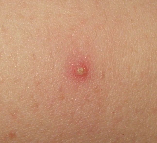
Folliculitis is infection or inflammation of the hair follicle, producing a pustule or red papule over hair-bearing skin. It is usually benign and self-limiting, but can progress. The usual sequence is folliculitis → furuncle → carbuncle: a furuncle (boil) is a deeper follicular abscess, and a carbuncle is a collection of two or more contiguous furuncles that spreads into the surrounding skin and subcutaneous tissue with multiple draining sinuses. Carbuncles favour the nape of the neck, back and thighs, often cause fever and malaise, and are linked with diabetes, obesity, immunodeficiency and MRSA colonisation.
| Type | Cause | Clue | Treatment |
|---|---|---|---|
| Superficial bacterial | S. aureus (MSSA or MRSA) | Commonest form | Often resolves alone; topical mupirocin or clindamycin; oral cephalexin or dicloxacillin if extensive or deeper |
| 'Hot tub' folliculitis | Pseudomonas aeruginosa (also Klebsiella, Enterobacter) | Contaminated pool or hot tub; follows prolonged oral antibiotics | Usually settles in 7–10 days; if needed, agents active against Pseudomonas such as ciprofloxacin |
| Pityrosporum (Malassezia) | Malassezia furfur (yeast) | Adolescents, shoulders, back and neck; acne worsens on antibiotics; KOH shows yeast | Oral itraconazole or fluconazole |
| Viral | Herpes simplex (rarely molluscum) | Grouped papulovesicles | Aciclovir, valaciclovir or famciclovir |
| Demodex | Demodex folliculorum mite | Controversial entity | Permethrin, ivermectin, metronidazole |
| Eosinophilic | Immune dysregulation, advanced HIV (low CD4) | Itchy follicular papules on scalp, face, neck; biopsy shows eosinophils | Treat underlying cause |
How are pyodermas asked in NEET PG and INI-CET?
- Organism: non-bullous impetigo — S. aureus (commonest) ± S. pyogenes; bullous impetigo — S. aureus.
- Image: honey-coloured crusts around the mouth, or flaccid bullae in a young child.
- Toxin and target: exfoliative toxin → desmoglein-1 → stratum granulosum in SSSS.
- Sequel: impetigo → post-streptococcal glomerulonephritis.
- Treatment: localised impetigo → topical mupirocin; extensive or bullous → systemic anti-staphylococcal antibiotic; MRSA → clindamycin or doxycycline.
- Compare: SSSS (Dsg1, mucosa spared) vs TEN (necrosis, mucosa involved) vs pemphigus vulgaris (Dsg3).