What are the boundaries of the popliteal fossa?
The popliteal fossa is a shallow diamond-shaped depression posterior to the knee joint, the knee 'pit'. It is a transit space for nerves, vessels and lymphatics between the thigh and the leg.
| Boundary | Formed by |
|---|---|
| Superomedial | Semimembranosus (and semitendinosus) |
| Superolateral | Biceps femoris (long and short heads) |
| Inferomedial | Medial head of gastrocnemius |
| Inferolateral | Lateral head of gastrocnemius, with plantaris running deep to it |
| Roof (superficial to deep) | Skin, superficial fascia, deep (popliteal) fascia |
| Floor | Popliteal surface of the femur, capsule of the knee joint with the popliteal ligament, and the fascia over popliteus |
What are the contents of the popliteal fossa and their order?
The tibial nerve is the most superficial structure of the neurovascular bundle, the popliteal vein lies superficial to the artery, and the popliteal artery is the deepest, lying next to the femur and knee capsule. The common peroneal nerve runs along the lateral boundary, beside the biceps femoris tendon.
| Structure | Key points |
|---|---|
| Tibial nerve | Branch of sciatic (L4–S3). Most superficial; crosses the fossa from superolateral to inferomedial. Gives muscular branches to gastrocnemius, soleus, plantaris and popliteus, articular branches, and the sural nerve (cutaneous) |
| Common peroneal (fibular) nerve | Branch of sciatic (L4–S2) at about the superior angle of the fossa; runs to the lateral angle and winds around the neck of the fibula, dividing into deep and superficial peroneal nerves. Gives the lateral sural cutaneous nerve; no direct motor branches in the fossa |
| Popliteal vein | Superficial to the artery. Formed at popliteus by the anterior tibial, posterior tibial and peroneal veins; receives the small saphenous vein, which pierces the deep fascia between the heads of gastrocnemius |
| Popliteal artery | Continuation of the femoral artery after the adductor hiatus; ends at the lower border of popliteus by dividing into the anterior tibial artery and the common trunk of posterior tibial and peroneal arteries. Deepest structure |
| Genicular arteries | Five branches: superior lateral, superior medial, middle, inferior lateral and inferior medial; they form a collateral network around the knee |
| Popliteal lymph nodes | In the fat and along the vessels; efferents mostly follow the femoral vessels to the deep inguinal nodes |
| Fat | Fills the space |
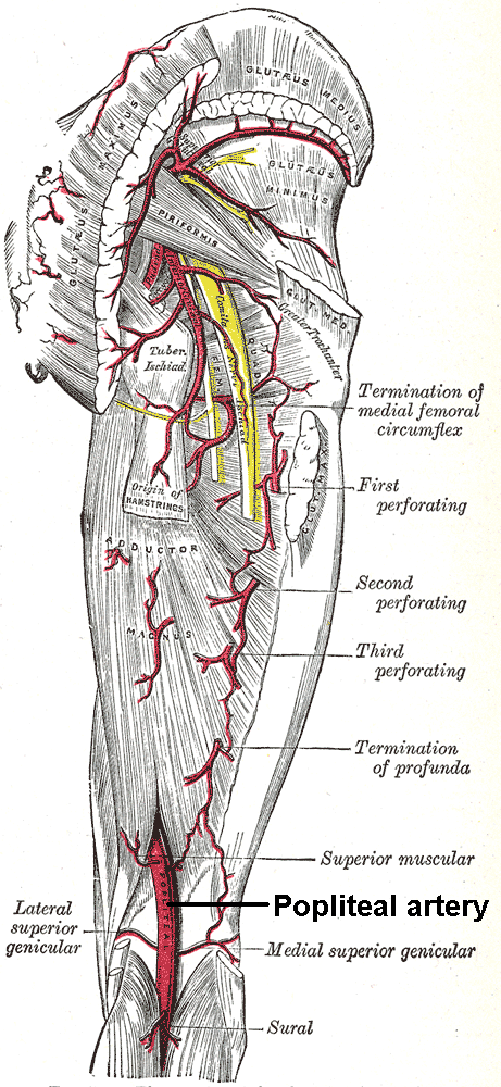
How is the popliteal pulse felt and what are the genicular anastomoses?
The popliteal pulse is deep and harder to feel than the radial or ankle pulses. Flex the knee slightly, grip the limb with both hands, thumbs over the patella and fingertips in the fossa. It is checked when the ankle pulses cannot be found, for example in peripheral arterial disease or diabetes.
The five genicular branches form a collateral network that links the femoral and popliteal arteries and the tibial arteries, giving an alternative route of blood flow around the knee. Superior genicular arteries connect to the deep femoral artery above, and inferior genicular arteries connect to the tibial arteries below. The middle genicular artery supplies the cruciate ligaments and synovial membrane. See also peripheral arterial disease and Buerger's disease.
What is a Baker cyst?
A Baker (popliteal) cyst is a fluid-filled sac behind the knee, typically between the semimembranosus tendon and the medial head of gastrocnemius. It is an enlargement of the gastrocnemius–semimembranosus bursa, which communicates with the knee joint in adults, so it is nearly always secondary to joint disease.
| Feature | Detail |
|---|---|
| Associations | Degenerative meniscal tears, osteoarthritis, rheumatoid arthritis and other inflammatory arthritis, infective arthritis, pigmented villonodular synovitis |
| Mechanism | Valve-like effect between joint and bursa: the meniscus or the gastrocnemius–semimembranosus junction lets synovial fluid pass in one direction |
| Age | Adults 35–70 years; in children it arises as a primary condition (herniated posterior capsule or synovium), most often at 4–7 years |
| Examination | Swelling most prominent with the knee fully extended; it softens or disappears at about 45° of flexion (Foucher sign) |
| Imaging | Ultrasound first; MRI if surgery is planned |
| Differentials | Popliteal artery aneurysm, DVT, ganglion, lipoma, haematoma, lymphadenopathy, sarcoma, abscess |

Complications. A large or ruptured cyst can dissect into the calf and mimic thrombophlebitis or DVT, with calf swelling, erythema, a positive Homans sign and a sensation of water running down the calf (pseudothrombophlebitis). It can also compress veins, entrap the posterior tibial nerve, occlude the popliteal artery, or cause anterior or posterior compartment syndrome.
Treatment. Asymptomatic cysts need only observation and reassurance. Symptomatic ones are managed with activity modification, NSAIDs, physiotherapy, and ultrasound-guided aspiration with steroid injection. Always treat the underlying joint disorder. Arthroscopic debridement or open excision (posterior approach) are options but recurrence is common, especially in older patients with degenerative disease.
What is a popliteal artery aneurysm?
Popliteal artery aneurysm (PAA) is the most common peripheral arterial aneurysm, about 70% of peripheral aneurysms, and second only to abdominal aortic aneurysm overall. It is a true aneurysm involving all layers of the wall. Aneurysmal dilatation means a diameter at least 1.5 times normal; with a normal artery of 0.5–1.1 cm, this is about 1.5 cm or more.
| Feature | Detail |
|---|---|
| Who | About 95% male |
| Associations | Bilateral (contralateral aneurysm in about 50–54%); coexisting abdominal aortic aneurysm in 36–51% |
| Presentation | Pulsatile popliteal mass (about 60%); up to 50% present with acute or chronic limb ischaemia; claudication, rest pain, ulceration or blue toe syndrome. A thrombosed aneurysm may not be pulsatile |
| Complication mechanism | Mural thrombus causes distal thromboembolism and may occlude the artery; repeated emboli occlude tibial vessels |
| Diagnosis | Duplex ultrasonography first line; CT or MR angiography if unavailable; arteriography before revascularisation for ALI |
| Repair | Symptomatic, including acute limb ischaemia, regardless of size; asymptomatic patent aneurysm 2 cm or larger |
| Technique | Saphenous vein graft preferred; thrombolysis if outflow is poor; fasciotomy if ischaemic for more than 4–6 hours or compartment syndrome |
Which nerve injuries occur around the popliteal fossa?
The sciatic nerve divides into the tibial and common peroneal nerves at about the superior angle of the fossa, though a higher division is the commonest variant (even in the pelvis).
| Feature | Tibial nerve | Common peroneal (fibular) nerve |
|---|---|---|
| Roots | L4–S3 | L4–S2 |
| Motor action | Plantarflexes and inverts the foot | Dorsiflexes and everts the foot |
| Sensation | Sole of foot; sural nerve to lower posterior leg and lateral foot | Dorsum of foot; lateral leg |
| Vulnerability | Most superficial structure of the neurovascular bundle, so it is met first in a posterior approach | Superficial over the fibular neck, protected only by skin, fat and fascia |
| Injury result | Loss of plantarflexion and inversion; sole numbness | Foot drop, loss of dorsiflexion and eversion, numb lateral leg and dorsum of foot |
Peroneal neuropathy is the most common mononeuropathy of the lower limb. Typical causes are a fibular neck fracture, tibiofibular or tibial plateau fracture, a tight cast or splint, and compartment syndrome. In foot drop the toes drag during the swing phase of gait. Piriformis syndrome (sciatic nerve entrapment) can mimic it, so check for symptoms above the knee, as in sciatica. Related pages: lower limb nerve injuries and gluteal region.