What are the features of molluscum contagiosum?
Molluscum contagiosum (MC) is a benign epidermal eruption caused by a double-stranded DNA virus of the Poxviridae family. Humans are the only known host, and the virus infects only epidermal keratinocytes; it replicates in the cytoplasm and cannot be cultured in standard cell lines. Lesions stay in the skin — there is no viraemia or systemic illness.
- Spread by skin-to-skin contact, autoinoculation (scratching) and fomites — towels, gym equipment, pool surfaces; in adolescents and adults also sexual contact.
- Incubation about 2–6 weeks (range from 1 week to 6 months); lesions usually resolve within a year.
- MCV-1 causes most paediatric cases; MCV-2 is commoner in sexually transmitted infection and in severe disease in the immunocompromised.
- Sites — trunk, limbs and face in children; genital, perineal and lower abdominal areas in sexually active adults. The oral mucosa is rarely involved.
- Children typically have 10–20 lesions; children with atopic dermatitis are at particular risk of extensive disease.
- Molluscum dermatitis (red scaly eczema around lesions) is an immune reaction and often a sign that lesions are about to resolve — not a bacterial infection.
- HIV / advanced immunodeficiency — numerous lesions, 'giant' molluscum (10–15 mm or more), atypical sites such as face and eyelids, and resistance to treatment.

How is molluscum contagiosum treated?
Observation ('benign neglect') is first-line in immunocompetent patients because most lesions clear spontaneously and no treatment is consistently superior. Treat when lesions are persistent, symptomatic, cosmetically distressing, or to reduce spread.
| Option | Details |
|---|---|
| Observation | First-line in immunocompetent children and adults; counsel about autoinoculation and fomites |
| Curettage | Often the most effective option, with good cosmetic results; preferred over electrodesiccation in children |
| Cryotherapy | Effective but painful in young children; may cause blistering and pigmentary change |
| Cantharidin 0.7% (VP-102) | Standardised single-use applicator, FDA-approved in 2020; complete clearance 46–54% vs 13–18% vehicle at 12 weeks |
| Berdazimer gel 10.3% | FDA-approved in 2024 for age 1 year and above; first at-home topical therapy, applied once daily |
| Potassium hydroxide, podophyllotoxin, salicylic or benzoyl peroxide | Variable efficacy and may irritate |
| Imiquimod | Not recommended — ineffective with more local reactions |
What are the types of viral warts and which HPV types cause them?
Warts are benign proliferations caused by HPV, a double-stranded DNA virus; over 150 HPV types are known, and only some infect skin. Infection begins in the basal layer of the epidermis, causing keratinocyte proliferation and hyperkeratosis. Spread is by skin contact or autoinoculation — scratching can produce a line of warts (pseudo-Koebner phenomenon). The incubation period can reach 12 months. Children, people with dermatitis (damaged barrier) and the immunosuppressed are most often affected.
| Type | Appearance and site | HPV types |
|---|---|---|
| Common wart (verruca vulgaris) | Cauliflower-like, rough, hyperkeratotic papule, 1 mm to 1 cm; knees, backs of fingers/toes, periungual | Skin types 1, 2, 3, 4, 10, 27, 29, 57 (the commonest skin types) |
| Plantar wart — myrmecia | Tender, deep wart on the sole with surrounding callus; painful on lateral pressure; skin lines interrupted | HPV 1 |
| Plantar wart — mosaic | Clusters of superficial, less painful warts | HPV 2 |
| Plane (flat) wart | Multiple flat-topped skin-coloured papules on face, hands, shins; spread by shaving | HPV 3 and 10 |
| Filiform wart | Finger-like fronds on a narrow stalk, usually on the face | — |
| Butcher's wart | Resembles common warts, numerous, on hands of those handling raw meat in cold, moist conditions | HPV 7 |

How are warts treated, and what about genital warts?
Most cutaneous warts resolve spontaneously: in children about 50% clear within six months and 90% within two years. Treat for immunosuppression, complications (nail damage, painful plantar warts) or patient preference. Treatments remove virus-containing skin; they do not kill the virus, so recurrence is common.
| Method | Key point |
|---|---|
| Salicylic acid or podophyllin paints, pastes, patches | Applied daily after paring; about 70% of warts resolve within 12 weeks |
| Cryotherapy (liquid nitrogen) | Repeat every 1–2 weeks; about 70% success after 3–4 months; blistering and possible white scar |
| Electrosurgery (curettage and cautery) | For large resistant warts; scarring; about 20% recur within months |
| Others | Imiquimod (approved for anogenital warts, usually ineffective on skin warts), bleomycin injection, pulsed-dye laser, contact immunotherapy such as diphencyprone or squaric acid |
- Genital warts (condylomata acuminata) — HPV 6 and 11 cause about 90%. Only 14 of the 200-plus HPV types are considered high-risk for malignant change (for example types 16 and 18), and they are different from the types in genital warts.
- Genital wart therapy includes imiquimod 5% cream three times weekly, trichloroacetic acid (clinic use) and physical removal.
- HPV vaccines prevent anogenital warts; see cervical cancer screening for the vaccine and screening context.
- Recurrence is commoner in smokers and the immunosuppressed; immunity is likely type-specific.
What are the clinical features of herpes simplex infection?
HSV is a double-stranded DNA herpesvirus. HSV-1 mainly causes oral and facial disease, HSV-2 mainly genital and rectal disease, though either can infect any skin or mucosa. After the primary episode the virus remains latent in the dorsal root ganglia and travels along the nerve to the skin during recurrence. A lesion is infectious for about 7–12 days, and the incubation period is 2–12 days.
| Presentation | Features |
|---|---|
| Primary HSV-1 | Often gingivostomatitis in children aged 1–5 — fever, drooling, swollen bleeding gums, vesicles becoming ulcers; settles in about 2 weeks |
| Primary HSV-2 | Genital herpes after onset of sexual activity — painful vesicles and ulcers for 2–3 weeks, fever, tender inguinal nodes |
| Recurrent | Itch or burning, then grouped, small, often umbilicated vesicles on a red base; heal in 7–10 days; triggers include fever, sun, trauma, stress and menstruation |
| Herpetic whitlow | Infection of the finger (health-care workers, thumb-suckers) |
| Eczema herpeticum | Severe widespread HSV in patients with atopic dermatitis or Darier disease |
- Complications — dendritic corneal ulcer, erythema multiforme (target lesions), facial nerve palsy, rarely meningitis; disseminated disease in immunocompromised patients.
- Diagnosis — clinical; confirm doubtful cases by PCR or culture of a swab from a fresh vesicle. Serology is of little use because most adults are positive.
- Treatment — mild disease needs none. Antivirals shorten attacks but cannot clear latent virus: aciclovir 200 mg five times daily for 5 days, valaciclovir 500 mg twice daily for 5 days, or famciclovir; longer or higher doses for immunocompromised patients or eczema herpeticum.
What is herpes zoster and how is it treated?
Herpes zoster (shingles) is reactivation of latent varicella-zoster virus from cranial nerve or dorsal root ganglia, driven by loss of cell-mediated immunity. Triggers include emotional stress, immunosuppressive drugs, illness and malignancy. Incidence is about 1.2–3.4 per 1000 person-years in healthy younger adults and 3.9–11.8 per 1000 in those over 65.
- Prodrome — burning pain, malaise, fever, 48 hours or more before the rash.
- Eruption — grouped vesicles on an erythematous base, in 1–3 crops over 3–5 days, unilateral and limited to one dermatome (thoracic about 53%, cervical 20%, trigeminal 15%, lumbosacral 11%). Patients are infectious until lesions crust.
- Herpes zoster ophthalmicus — ophthalmic division of the trigeminal nerve, in roughly 10–25% of cases; Hutchinson sign (vesicles on the nasal tip) points to nasociliary involvement and a high risk of ocular disease.
- Ramsay Hunt syndrome — zoster of the facial and vestibulocochlear nerves causing ear vesicles, hearing loss and vertigo.
- Postherpetic neuralgia — pain lasting beyond about 4 weeks, sometimes 12 months or more; the principal morbidity.
| Drug | Dose |
|---|---|
| Aciclovir | 800 mg, five times daily for 5 days |
| Valaciclovir | 1 g three times daily for 5 days |
| Famciclovir | 500 mg three times daily for 7 days (alternative if aciclovir resistance) |
| IV aciclovir (immunocompromised) | 10 mg/kg every 8 hours for 7 days; dilute and infuse slowly to avoid crystal nephropathy |
| Neuropathic pain | Gabapentin or pregabalin first line; tricyclics also used; lidocaine 5% or capsaicin 8% patches |
What does a Tzanck smear show, and how are these infections confirmed?
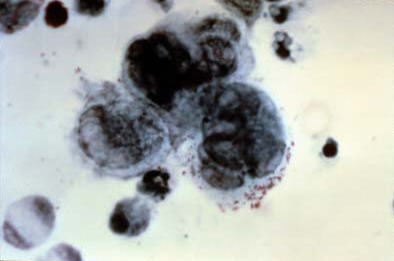
A Tzanck smear is a stained scraping from the base of a fresh vesicle. In varicella-zoster infection it shows multinucleated giant cells. It is quick but has lower sensitivity and specificity than direct fluorescent antibody testing or PCR, which are preferred when confirmation is needed.
| Infection | Confirmatory test | Histology / cytology clue |
|---|---|---|
| Molluscum | Clinical; biopsy or cytology if atypical; PCR (p43K gene) | Henderson-Paterson bodies |
| Warts | Clinical; paring shows capillary dots; biopsy if squamous cell carcinoma is possible | Papillomatosis, hyperkeratosis |
| HSV | PCR or culture of fresh vesicle swab | — |
| Zoster | Clinical; Tzanck, DFA or PCR; VZV IgM during active infection | Multinucleated giant cells |