What is the lumbosacral plexus and how is it formed?
StatPearls defines the lumbosacral plexus as a network formed by the anterior rami of the L1 to S4 nerve roots, with a small contribution from T12. It is the combination of two plexuses. The lumbar plexus (L1 to L4) lies above the pelvic brim; the sacral plexus (S1 to S4) lies below the pelvic brim. The two are joined by the lumbosacral trunk, which carries L4 (its lower part) and L5 down into the pelvis.
The anterior rami of the first four lumbar nerves descend through the psoas major and each splits into anterior and posterior divisions. These divisions recombine into the named nerves. The lumbar plexus is embedded in the posterior part of psoas, so its blood supply is shared with the muscle — chiefly the lumbar branch of the iliolumbar artery — and ischaemic injury during aortoiliac surgery is uncommon.
| Feature | Lumbar plexus | Sacral plexus |
|---|---|---|
| Roots | L1 to L4 (T12 contributes) | L4/L5 (via lumbosacral trunk) and S1 to S4 |
| Position | Inside psoas major, above the pelvic brim | On the pelvic wall, below the brim, in front of piriformis |
| Largest nerve | Femoral (posterior divisions of L2 to L4) | Sciatic (L4 to S3), the largest nerve in the body |
| Main territory | Lower abdominal wall, anterior and medial thigh | Gluteal region, posterior thigh, leg, foot, perineum |

What are the branches of the lumbar plexus and their root values?
StatPearls lists the nerves of the lumbar plexus from superior to inferior as the iliohypogastric, ilioinguinal, genitofemoral, lateral femoral cutaneous, femoral and obturator nerves, plus the contribution to the lumbosacral trunk. Short direct branches also supply the psoas, quadratus lumborum and lumbar transverse muscles.
| Nerve | Roots | Motor | Sensory |
|---|---|---|---|
| Iliohypogastric | T12, L1 | Internal oblique, transversus abdominis | Lateral gluteal skin |
| Ilioinguinal | L1 | Internal oblique, transversus abdominis | Upper medial thigh; root of penis and anterior scrotum, or mons pubis and labia majora |
| Genitofemoral | L1, L2 | Genital branch: cremaster | Genital branch: scrotal skin or mons/labia majora; femoral branch: skin of anterior upper thigh |
| Lateral femoral cutaneous | L2, L3 | None (purely sensory) | Lateral thigh |
| Femoral | L2, L3, L4 (posterior divisions) | Quadriceps, sartorius, articularis genu; iliacus and pectineus | Anterior thigh; saphenous nerve to medial leg and foot |
| Obturator | L2, L3, L4 (anterior divisions) | External obturator, adductor longus, brevis and magnus, gracilis, pectineus | Medial thigh (below the ilioinguinal territory) |
The femoral nerve is the largest nerve of the lumbar plexus. It enters the femoral triangle lateral to the femoral artery and has four types of branch: muscular, cutaneous, articular and vascular. Its saphenous branch supplies skin over the patella and the medial and anterior side of the distal leg. The obturator nerve is the main motor supply of the medial (adductor) compartment and also sends articular branches to the hip and knee.
What are the branches of the sacral plexus and their root values?
The sacral plexus is primarily composed of the superior gluteal (L4 to S1), inferior gluteal (L5 to S2), posterior femoral cutaneous (S1 to S3), sciatic (L4 to S3) and pudendal nerves. Divisions matter here too: the sciatic nerve carries fibres from both anterior and posterior divisions, which is why it later splits into tibial (anterior-division fibres) and common fibular (posterior-division fibres) parts.
| Nerve | Roots | Exit from pelvis | Supply |
|---|---|---|---|
| Superior gluteal | L4, L5, S1 (dorsal divisions) | Greater sciatic foramen, above piriformis | Gluteus medius, gluteus minimus, tensor fasciae latae |
| Inferior gluteal | L5, S1, S2 | Greater sciatic foramen, below piriformis | Gluteus maximus (motor only) |
| Sciatic | L4 to S3 | Greater sciatic foramen, below piriformis | Posterior thigh; all muscles below the knee via tibial and common fibular nerves |
| Posterior femoral cutaneous | S1, S2, S3 | Below piriformis, with the sciatic nerve | Skin of the posterior thigh |
| Pudendal | S2, S3, S4 | Greater sciatic foramen, then re-enters by the lesser sciatic foramen | Perineum, external anal and urethral sphincters, external genitalia |
| Nerve to obturator internus | Sacral plexus | Below piriformis | Obturator internus |
The sciatic nerve leaves the pelvis through the greater sciatic foramen inferior to piriformis, together with the pudendal nerve and vessels, inferior gluteal nerve and vessels, nerve to obturator internus and posterior femoral cutaneous nerve. The superior gluteal nerve is the one structure that exits above piriformis, accompanied by the superior gluteal artery and vein.

What are the signs of sciatic, gluteal and fibular nerve injury?
The sciatic nerve innervates the hamstrings (biceps femoris, semitendinosus, semimembranosus and the ischial part of adductor magnus). The tibial division supplies the posterior leg and foot (plantar flexion, toe flexion); the common fibular division supplies the anterior and lateral compartments (dorsiflexion, toe extension and eversion). A high sciatic lesion therefore weakens knee flexion and every movement below the knee.
| Nerve | Deficit | Typical clue |
|---|---|---|
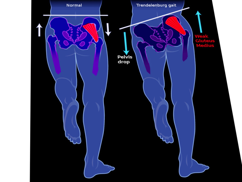
| Superior gluteal | Weak hip abduction; positive Trendelenburg sign | Pelvis sags on the normal (swing) side when standing on the affected leg; injured by hip dislocation or a badly placed intramuscular injection |
| Inferior gluteal | Gluteus maximus weakness and atrophy; difficulty rising from sitting, climbing stairs | Entrapment below piriformis; extension of the trunk from a forward bend is reduced |
| Sciatic (piriformis syndrome) | Buttock pain radiating down the limb, worse on sitting and driving | Swollen piriformis compresses the nerve; no paralumbar spasm, unlike disc prolapse |
| Common fibular | Foot drop, loss of eversion, sensory loss on the dorsum of the foot | Most common compressive neuropathy of the lower limb; fibular-neck compression or knee dislocation |
In a normal gait the small gluteal muscles on the standing leg stabilise the pelvis in the coronal plane. If the superior gluteal nerve is damaged, the opposite pelvis is not lifted and sags: the Trendelenburg sign. Bilateral damage gives a waddling (gluteal) gait.
What are the signs of femoral, obturator and cutaneous nerve injury?
| Nerve | Deficit | Typical cause or clue |
|---|---|---|
| Femoral | Quadriceps wasting, loss of knee extension, weaker hip flexion; sensory loss over anterior and medial thigh (and saphenous territory) | Psoas or iliacus haematoma or abscess, hip surgery, lithotomy position at vaginal delivery compressing the nerve against the inguinal ligament |
| Obturator | Weak thigh adduction; medial thigh sensory loss | Pelvic surgery, obturator hernia, entrapment in athletes; stimulation causes the adductor jerk during transurethral bladder tumour resection |
| Lateral femoral cutaneous | Burning pain and numbness on the lateral thigh, no weakness | Meralgia paraesthetica: entrapment under or through the inguinal ligament |
| Genitofemoral | Groin and upper thigh pain or sensory change; cremasteric reflex lost | Hernia repair, groin trauma, saphenous vein surgery retractors |
In femoral nerve injury a patient may fall because the quadriceps no longer works; the patellar tendon reflex (L4) can be lost when the L4 root is affected. L4 disc herniation can therefore mimic femoral neuropathy: the clue to a radiculopathy is dermatomal pain and involvement of muscles outside the femoral territory.
The cremasteric reflex: stroking the medial thigh makes the cremaster contract and raises the testis. The afferent limb is the femoral branch of the genitofemoral nerve (with ilioinguinal), the efferent limb is the genital branch. StatPearls notes the reflex is absent in testicular torsion.
What is the course of the pudendal nerve and why does it matter?
The pudendal nerve carries motor and sensory fibres from the ventral rami of S2 to S4. It leaves the pelvis by the greater sciatic foramen, passes through the gluteal region proximal to the ischial spine, wraps around the sacrospinous ligament and enters the perineum through the lesser sciatic foramen. It then runs in the pudendal (Alcock's) canal on the medial wall of obturator internus.
- Inferior rectal (anal) nerve — motor to the external anal sphincter; carries sensation from the anal canal below the pectinate line.
- Perineal nerve — the middle branch, to the perineum.
- Dorsal nerve of the penis or clitoris — sensation of the penis or clitoris.
The pudendal nerve also supplies the lower fifth of the vagina. Because it sits next to the ischial spine, a pudendal nerve block at that point gives analgesia in labour. The nerve is vulnerable to stretch injury during childbirth, especially with a heavy baby; fixed injury can cause faecal or urinary incontinence, chronic pain or sexual dysfunction.
What are the causes and features of lumbosacral plexopathy?
Lumbosacral plexopathy is an injury of the lumbar and sacral plexus nerves. It is far less common than brachial plexopathy and can be hard to diagnose. Patients present with low back and leg pain, motor weakness, numbness or paraesthesia; sphincter dysfunction is uncommon and, if present, suggests cauda equina syndrome.
| Group | Examples |
|---|---|
| Trauma | Posterior hip dislocation, sacral fracture, pelvic fracture, penetrating injury |
| Metabolic or immune | Diabetic amyotrophy (diabetic lumbosacral radiculoplexus neuropathy), amyloidosis, sarcoidosis |
| Neoplastic | Compression or invasion by abdominopelvic malignancy (more than 70 percent of neoplastic cases) |
| Radiation | Delayed fibrosis and microvascular ischaemia; often painless |
| Obstetric and haematoma | Birth trauma in late pregnancy or after delivery; retroperitoneal haematoma after femoral catheterisation or pelvic surgery |
| Infection | Psoas abscess, tuberculosis, vertebral osteomyelitis, herpes zoster |
Diabetic amyotrophy typically causes unilateral proximal thigh pain, then weakness and wasting, mostly in type 2 diabetes. StatPearls gives an incidence of 4.2 per 100,000 per year, affecting 0.8 percent of people with diabetes. Neoplastic plexopathy mostly involves the L4 to S1 segments. Electromyography helps localise the lesion; MRI or CT evaluates tumour, haematoma or abscess.
What should I memorise for the exam?
- Lumbar plexus lies in psoas major; sacral plexus lies on the pelvic wall in front of piriformis.
- Femoral = posterior divisions L2–L4; obturator = anterior divisions L2–L4.
- Lateral femoral cutaneous is L2–L3, purely sensory; meralgia paraesthetica = its entrapment.
- Genitofemoral L1–L2: genital branch is the motor and efferent limb of the cremasteric reflex.
- Superior gluteal (L4–S1) is the only branch exiting above piriformis; lesion = Trendelenburg sign.
- Inferior gluteal (L5–S2) supplies only gluteus maximus.
- Sciatic (L4–S3) leaves below piriformis; piriformis syndrome = sciatic compression.
- Pudendal S2–S4: exits greater, re-enters lesser sciatic foramen; Alcock's canal; block at the ischial spine.