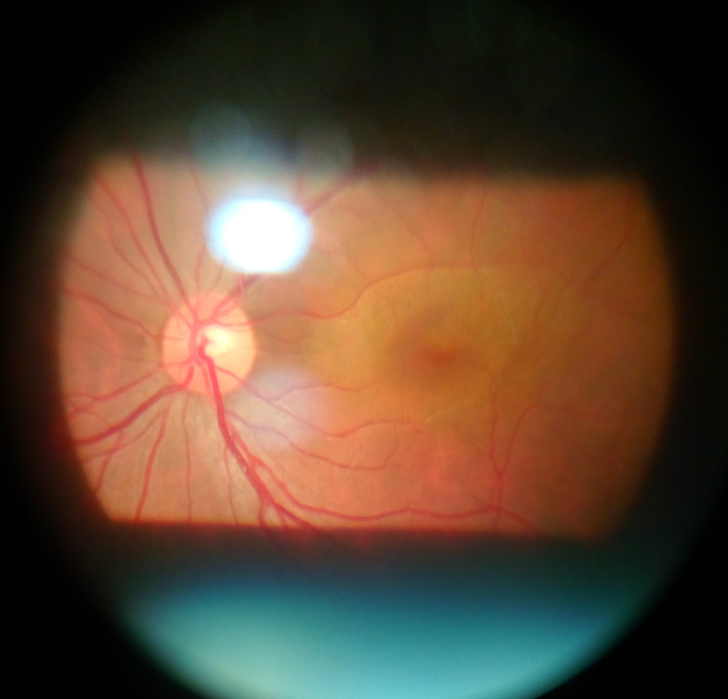
A middle-aged woman presents with pain on eye movement. There is a relative afferent pupillary defect (RAPD) and a normal optic disc on fundus examination. What is the most likely diagnosis?
- A.CRAO
- B.Retrobulbar optic neuritis
- C.Papilledema
- D.Anterior ischemic optic neuropathy
Show answer and explanation
Answer: B. Retrobulbar optic neuritis
The correct answer is Retrobulbar optic neuritis.
The triad of pain on eye movement, a relative afferent pupillary defect (RAPD), and a normal optic disc is classic for retrobulbar optic neuritis. The inflammation lies behind the globe, so the disc looks normal initially - hence the saying that the patient sees nothing and the doctor sees nothing. It is most often demyelinating (multiple sclerosis) in a young to middle-aged woman.
The closest distractor is anterior ischemic optic neuropathy (or papillitis). However, AION and papilledema both show an ABNORMAL (swollen) disc, and CRAO gives painless sudden vision loss with a cherry-red spot and pale retina. Only retrobulbar optic neuritis combines pain on eye movement, RAPD, and a normal disc.
Incorrect Options:
- CRAO: Sudden PAINLESS vision loss with a cherry-red spot and pale retina, not pain on movement.
- Papilledema: Bilateral disc SWELLING from raised intracranial pressure, with a swollen disc and usually no RAPD.
- Anterior ischemic optic neuropathy: Sudden painless loss with a SWOLLEN or pale disc, typically in older patients.
High-Yield Facts:
- Retrobulbar optic neuritis: pain on eye movement, RAPD, normal disc.
- Strongly associated with multiple sclerosis.
- Papillitis (anterior optic neuritis) shows a swollen disc.
- RAPD indicates an asymmetric optic nerve (afferent) lesion.
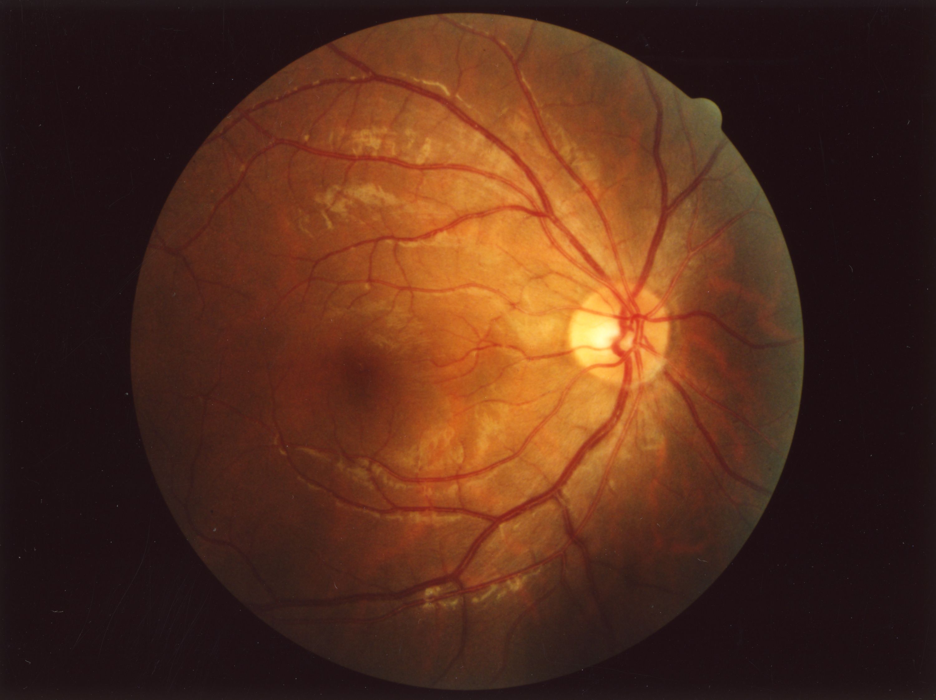
Figure: A normal-looking optic disc. In retrobulbar optic neuritis the disc appears normal because the inflammation is behind the globe.